7 Simple Techniques For Babyecho
7 Simple Techniques For Babyecho
Blog Article
The Best Guide To Babyecho
Table of ContentsSome Known Facts About Babyecho.Babyecho for DummiesAn Unbiased View of BabyechoThe smart Trick of Babyecho That Nobody is DiscussingThe 3-Minute Rule for BabyechoBabyecho Things To Know Before You BuyUnknown Facts About BabyechoThe 5-Second Trick For BabyechoThe 8-Minute Rule for Babyecho
You might not hear the infant's heart beat at the first trimester ultrasound visit, and that's alright. It may be due to the fact that you're not much sufficient along in the maternity or the infant's setting. In some cases the child's heartbeat can be detected as early as 5 to 6 weeks after perception. You can typically hear the heartbeat much better more detailed to 10 weeks after gestation.
Not known Incorrect Statements About Babyecho
Your doctor may recommend that you have 1 or even more ultrasounds at various points in your maternity - fetal heart doppler. Depending on just how much along your maternity is, ultrasound pictures aid your doctor: approximate your due date check things like the size and position of the fetus to make sure whatever is regular see the setting of the placenta see the quantity of amniotic fluid in your womb discover multiple maternities (doubles, triplets, etc) Ultrasounds can additionally be used to screen for particular birth defects, like Down syndrome. https://pastebin.com/u/babydoppler1.
Physicians, midwives, or educated ultrasound service technicians will do your ultrasound and read the outcomes. The price of an ultrasound depends on the kind of ultrasound you get and where you obtain it.
Unknown Facts About Babyecho
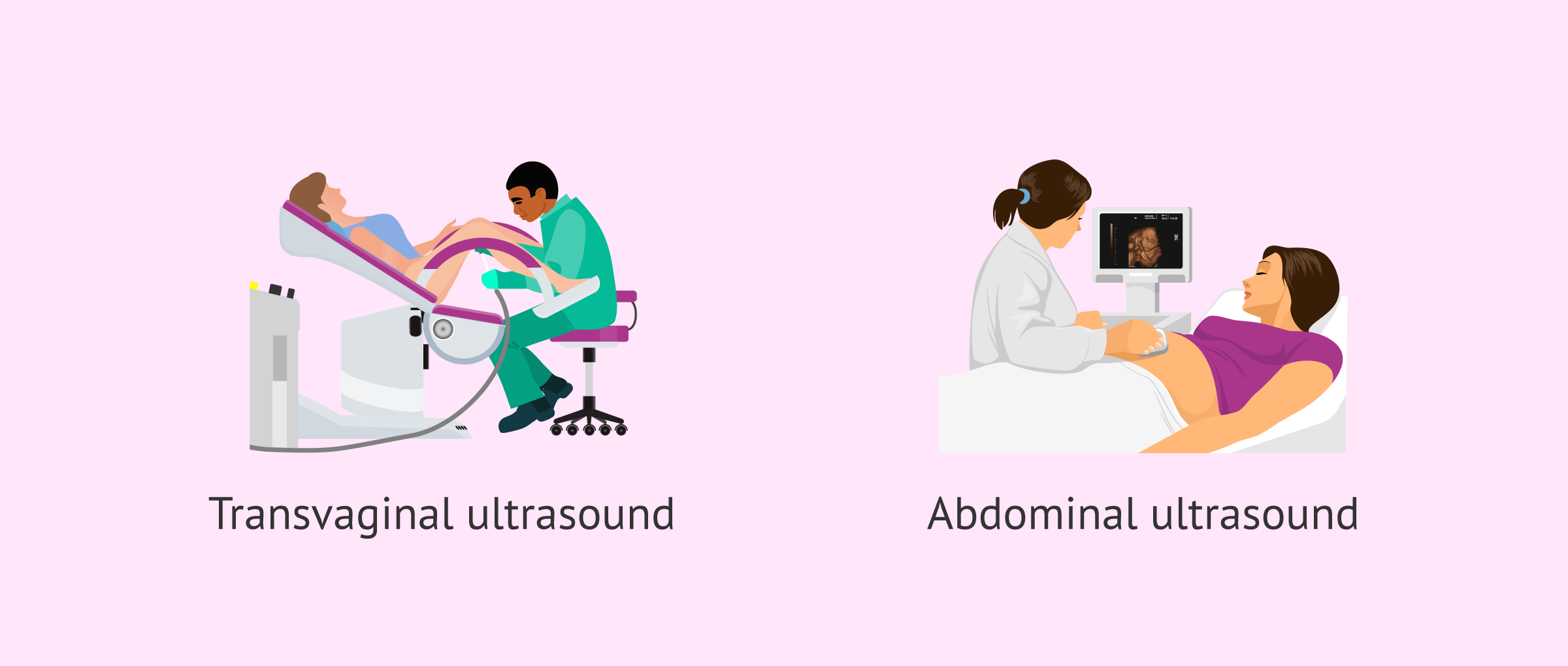
Ultrasound scans make use of sound waves to develop an image of your child on a TV screen. The scans are risk-free and can be performed at any kind of stage of your maternity. Most ultrasounds are 'transabdominal ultrasounds'. This implies that the ultrasound probe is massaged carefully on your belly (abdominal area) to produce a photo of the infant on the screen.
You will certainly be brought right into a darkened area. The darkness makes it less complicated for the person doing the check to plainly see the image on the display. You will be asked to rest on a sofa and raise up your top to reveal your stomach. You may need to roll down the waistband of your pants.
Examine This Report about Babyecho
This is to shield your garments from the ultrasound gel. Next, they will certainly put the ultrasound gel onto your stomach.
You may not be able to tell where your baby is in the picture. The person doing the check will typically point things out to you like the infant's heartbeat and head.
Not known Facts About Babyecho
It can be distressing when your scan suggests that there is a problem with your pregnancy or your baby. Your midwife, obstetrician and General practitioner are there to support you.
They can be pricey and you will certainly require to schedule the check yourself. Your GP, midwife or obstetrician will tell you where you can get personal maternity scans in your area. If a worry is located on a personal check, follow-on solutions might not be offered there. You will certainly require to contact your maternal medical facility.
Fetal ultrasound is a test done during maternity that uses shown audio waves. The picture is shown on a Television display. It may be in black and white or in colour.
Babyecho Things To Know Before You Get This
It can be done as early as the 5th week of maternity. Occasionally the sex of your fetus can be seen by regarding the 18th week of pregnancy.
Price quote the age of the unborn child (gestational age). Price quote the threat of a chromosome defect, such as Down syndrome. Examine for birth issues that impact the mind or spine. This examination is done to: Quote the age of the unborn child. Take a look at the dimension and setting of the unborn child, placenta, and amniotic fluid.
The Greatest Guide To Babyecho
It can be distressing when your scan check my reference recommends that there is a trouble with your pregnancy or your child. Your midwife, obstetrician and general practitioner are there to sustain you. Ask them to clarify whatever to you in information. You might require extra scans if: you have bleedingyou have bother with your child's movementsyour baby's growth requires to be monitoredExtra scans might additionally be needed to inspect: the position your infant is existing inthe position of the placenta (afterbirth)the amount of amniotic liquid around your babyMost pregnancy healthcare facilities do not regularly do scans to determine the sex of your infant.
:max_bytes(150000):strip_icc()/JoseLuisPelaezInc-17f79a53211940c2bc62cf23bc4185d4.jpg)
What Does Babyecho Do?
Fetal ultrasound is a test done during maternity that makes use of mirrored sound waves. It creates a photo of the infant (unborn child), the body organ that sustains the fetus (placenta), and the fluid that surrounds the unborn child (amniotic liquid). The picture is shown on a TV display. It may be in black and white or in colour.
It does not make use of X-rays or various other kinds of radiation that may damage your fetus. It can be done as early as the fifth week of maternity. In some cases the sex of your fetus can be seen by regarding the 18th week of pregnancy. Ultrasound is just one of the testing examinations that might be done in the very first trimester to search for abnormality, such as Down syndrome.
Getting The Babyecho To Work
Examine for birth defects that affect the mind or back cord. This examination is done to: Estimate the age of the unborn child. Look at the size and position of the unborn child, placenta, and amniotic fluid - doppler.
Report this page